Product recommendations · hot products
Rich experience and strong strength

100pcs histology microscope prepared slides

Source supplier with guaranteed quality

100% factory

More than 4000 types

Well-distributed coloration

Clearly cell tissue

Medical parasitology prepared slides set

Source supplier with guaranteed quality

Professional production

OEM & ODM

High-quality glass slides

Structure clear

85CM Sexless Torso 20 Parts Model

Source supplier with guaranteed quality

Non toxic safe materials

High product stability

Durable and sturdy

Exquisite details

Product Center ·Be an honest merchant
Rich experience and strong strength
Microscope Prepared Slides
Medical Teaching Models
Nursing Training Model
Histological Microtome
Microscope
Laboratory Consumables

Four Reasons to Choose Us

One stop procurement is more reassuring!

01、Quality certification and after-sales service

Quality Certification · Quality After Sales

We provide after-sales telephone technical support for you 7x24 hours We provide comprehensive services based on product requirements, performance, and after-sales service.

02、Strict standards and layer by layer testing

Strict production standards and reliable quality

Adopting a strict quality management system as the standard, continuously optimizing management. Products must go through strict control from material selection, processing, output to quality inspection!

03、Continuous Innovation · Service Market

Adhere to innovative development

Adhere to a customer-oriented product development and innovation philosophy. With years of industry experience, we have served over 2000 clients in total!

04、 Powerful manufacturer · spot direct sales

Source factory, one-stop service provider

Our products are directly sold by manufacturers with guaranteed quality The product has sufficient supply and direct sales in stock, supporting customized production on demand!
Tel:+86 18337360216
direct dealkeep a large stock of goods
Rich experience and strong strength

About us

Be an honest merchant

Xinxiang Vic Science&Education Co.,Ltd.

Founded in 2014, Xinxiang Vic Science&Education Co.,Ltd. is located in Hi-Tech Zone, Xinxiang city near to the Yellow River and Zhengzhou Airport. Specializing in production of microscope prepared......
Learn morethe news
Rich experience and strong strength

The Application of Immersion Specimen in Teaching

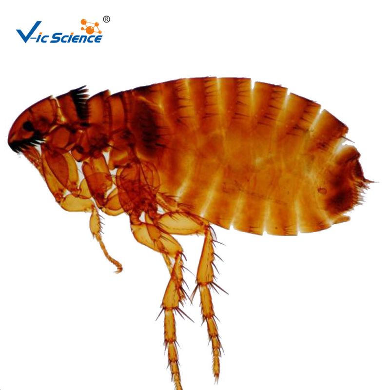
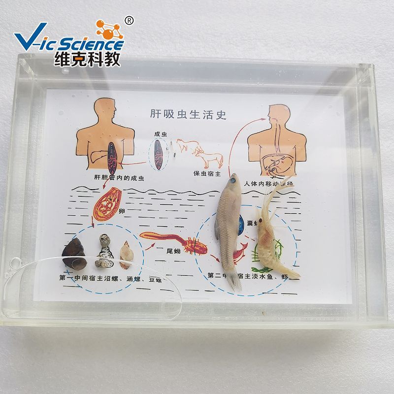
Immersion specimens have various applications in teaching:1、 Visual display of biological morphology and structurePlant teaching: In botany teaching, soaking specimens can effectively showcase the organs of plants. For example, when displaying immersed specimens of flowers, students can clearly see the number, shape, and color of petals, as well as the structure of stamens (including stamens and pistils). Soak specimens like lilies allow students to observe that they have 6 petals, 6 stamens, and 1 pistil, and can be stored for a long time, making it convenient for students to observe at any time without being limited by the flowering period. For the fruits and seeds of plants, soaking specimens can also display their external morphology and internal structure. For example, by soaking pea pod specimens, students can see the arrangement of pea seeds in the pods.Animal teaching: In animal science teaching, specimen immersion allows students to intuitively understand the appearance of animals. Taking fish as an example, immersion specimens can display the fish's body size, types of fins (dorsal fin, pectoral fin, pelvic fin, anal fin, and caudal fin), and their positions. Students can observe the morphological differences of fins in different fish species, such as the elongated dorsal fin of crucian carp and the wider dorsal fin of fighting fish. For some small invertebrates, such as earthworm specimens, their link structures can be clearly presented, helping students understand the characteristics of link animals.2、 Assist in explaining knowledge of biological classificationClassification display: Immersion specimens can serve as physical display materials for biological classification. For example, when explaining the classification of vertebrates, display immersion specimens from different classes (such as fish, amphibians, reptiles, birds, and mammals). By comparing the immersion specimens of fish (with gill respiration and fins) and amphibians (with gill respiration for juveniles, lung respiration and skin assisted respiration for adults, and limbs), students can more intuitively understand the main differences between different classes and master the basic knowledge of biological classification.Species diversity presentation: Teachers can use immersion specimens to display species diversity within a specific group. For example, when explaining the insect class, display immersion specimens of various insects such as locusts, butterflies, bees, etc. Students can observe differences in wing morphology (locusts have leathery forewings and membranous hindwings, butterflies have colorful and membranous wings) and mouthparts types (locusts have chewing mouthparts, butterflies have siphon mouthparts), and appreciate the richness and diversity of insect species.3、Help understand the ecological relationships of organismsFood chain display: A simple food chain model can be constructed for teaching through a series of immersion specimens. For example, by displaying immersion specimens of plankton, small fish, and large fish, teachers can explain the relationship between plankton as primary producers being preyed upon by small fish and small fish being preyed upon by large fish, allowing students to better understand the energy flow in the food chain and ecosystem.Biological habitat environment correlation: Immersion specimens can also be combined with an introduction to the biological habitat environment. For example, when displaying immersion specimens related to mangrove ecosystems, immersion specimens such as mangrove plants (such as okra), fiddler crabs, and mudskippers living in mangroves are also displayed, accompanied by pictures or videos of the muddy intertidal environment of mangroves, allowing students to understand the interrelationships between these organisms and their environment.
+ 2025-07-01

Advantages of tissue microtome

Tissue microtome is a kind of equipment for pathological section analysis of human tissues, animal and plant tissues. Tissue microtome are generally divided into rotary microtome and frozen microtome. Rotary microtome: It is used to slice by rotating the hand wheel. The wax block table is embedded in the metal clamp seat that can move up and down in the groove, and cuts the flat slice by pushing forward with the micro screw. Some rotary microtome are equipped with three knobs and a fastening knob on the head, which can deflect and tighten it in all directions, so as to adjust the section of the wax block. The cutting angle of the slicer can be adjusted. Because this microtome uses a heavy and large slicer knife, it generally does not vibrate when cutting hard tissue. The slice thickness can be adjusted from 1-30 microns to any thickness by the knob, and each gradient is 1 or 2 microns. The advantage of the rotary microtome is that the body is heavy, so it is more stable than the rocking microtome. It is very suitable for cutting paraffin sections. It can be ideal for cutting continuous sections and also for cutting large tissue blocks. 
+ 2024-06-06

06

2024-06

Paraffin tissue section preservation

Many laboratory researchers are accustomed to storing tissue wax blocks for a long time at room temperature after sectioning, and the tissue after sectioning is prone to antigen loss for long-term storage at room temperature.  Some studies have found that when sections are stored at room temperature for more than 3 months, immunohistochemical staining results are weakened or even negative. Therefore, some scholars have carried out new and old slice preservation time control experiments. 11 different tissue wax blocks were selected and 10 pieces were cut continuously for a total of 110 pieces per case, and placed in room temperature air for 1 month, 3 months, 6 months, and 1 year, and then the control experiment was carried out with new sections. The experimental results showed that when stored at room temperature for 3 months after wax sectioning, the sensitivity of antigens to most antibodies decreased by 50%, and the antigen loss of some sections was more. Most antibodies did not show positive results after 6 months of sectional storage, and only a few sections had weak positive expression after storage for more than 1 year, especially nuclear expression antigen loss was more prominent.  The reason may be related to oxidation, so in practical work, wax sealing sections and -20 degrees cryopreservation tissue sections can be used to avoid antigen loss, or paraffin tissue blocks can be stored at 4 degrees, and then sectioned when used, which can greatly reduce the antigen loss rate.

06

2024-06

The production process of frozen sections

Frozen sectioning method is the most commonly used sectioning technique in histochemistry, especially enzyme histochemistry, fixed or unfixed tissue samples can be frozen section, fresh tissue frozen sections for tissue intracellular enzyme preservation is also a commonly used sectioning method in immunocytochemical research, can better preserve a variety of antigens, especially the antigenicity of surface antigens.  1. PreparationClean up the test bench and instrumentsTurn on the cryomicrotome and pre-reduce the cryomicrotome to the required temperature (-15~-20 °C)Prepare the processed slides, blades, glue and medicines  2. Clamp the frozen tissue block on the microtome holder, start the rough advance and withdrawal button, turn the knob, and smooth the tissue. 3. Adjust the thickness to be cut, according to different tissues, in principle, it is a cell-intensive thin cut, and the fiber multicellular thin can be slightly thick cut, generally between 5 ~ 10μm. 4. Adjust the anti-coil plate. The key to making frozen slices lies in the adjustment of the anti-roll plate, which requires the operator to be careful and accurately adjust it well and adjust it to the appropriate position. When slicing, cut out a complete, smooth slice. When the cut tissue adheres to a clean slide, gently apply a gentle force in one direction to avoid wrinkles during the tissue spreading process, ensure the integrity of the tissue structure and the beauty of the section.
HOME TEL TOP